The New Technologies Accelerating Cell Line Development
Explore how AI, automation and gene-editing tools are reshaping cell line development to speed up and simplify biologic drug production.
At the turn of the 21st century, biologics – drugs derived from biological sources – were a small peak in the drug development landscape. This broad category of drugs includes monoclonal antibodies, enzymes, cytokines and vaccines, but in 2000, just 7% of US Food and Drug Administration (FDA) approvals were biologics. Instead, small-molecule drugs dominated the market. Today, things have changed drastically. A robust 40–60 split between biologics and small-molecule drugs has become the norm.1
The evolution of cell line development
To achieve this progress, biologic production has had to reinvent itself. Although biologics were once sourced directly from donated blood plasma or cells, the backbone of this process is now a structured, reproducible approach known as cell line development (CLD). Chinese hamster ovary (CHO) cells are the workhorse of modern CLD, and most biologics are synthesized using engineered CHO lines.2
Mammalian cell lines, such as CHO, have been widely adopted due to their reliable growth patterns in cell-free media and their ability to produce molecules with human-compatible glycosylation at high titers.
Nonetheless, CHO has a significantly longer doubling time than other host cell types, such as yeast or bacteria, making accuracy and speed paramount. In response, automation and digitization of processes have become widespread within the field.
Traditional CLD included several steps that involved laborious manual work. CLD workflows generally begin with the development of expression plasmids, which are then transfected into host cells.
Next, selection steps isolate cells that have been successfully and robustly transfected. Single cells with high productivity must be isolated and cultivated. These are then clonally expanded and assessed for protein expression. The top performers then undergo further testing ahead of scaling up. Without automation, this process can take six to nine months of testing.3
The most time-consuming processes are the single-cell cloning, monoclonality assurance and clone screening steps, which each take several weeks. The need for sterility, as well as the challenges of identifying and isolating individual cells in each well and of linked screening steps, further hinder the process. Proof of uniformity of cell lines in the biologics production process is a key requirement of regulatory agencies such as the FDA. With these high stakes, automation can streamline and accelerate the process to enhance accuracy.
Streamlining single-cell cloning
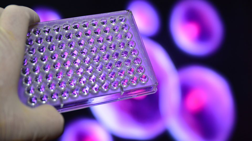
The strict requirements to show a monoclonal origin for biologics mean that, after transfection, CLD processes must isolate single cells into 96-well plates. Before automation, this was a mighty task. “Without any automation – what we have right now – many of the cell cloning steps are really based on limited dilution,” said Fang Tian, director of biological content at non-profit biological resource ATCC.
Limited dilution uses a statistical nutcracker to break this complex task. Transfected cell populations are serially diluted down to a level where individual wells are likely to only contain a single cell. This technique is gentle on cells, improving viability; however, the statistical distribution that predicts the likelihood of only a single cell being present limits the process’s efficiency.
At a seeding density of 1 cell/well, roughly a third of wells should contain a single cell.4 This is an inefficient success ratio that may necessitate a time-consuming additional round of limited dilution. “A single clone is vital,” said Tian. “When you have a mixture of clones, then some clones will overgrow and take over the entire population.”
A High Productivity CHO Expression System for Continuous Antibody Manufacturing
Discover how to achieve exceptionally high antibody titers.View Webinar
Luckily, automation has proved adept at modernizing this process. Liquid handlers and fluorescence-activated cell sorters (FACS) can distribute single cells across up to 384-well plates. Multiple studies have shown that the adoption of these systems can significantly reduce development time and the number of screening assays.5,6
Two drawbacks of the original generation of FACS machines are the high pressure they subject cells to, which can reduce viability, and the risk of cross-contamination between samples.
The latest generation of these machines utilizes microfluidics technologies to relieve pressure and maintain cell survival. Disposable cartridges maintain sterility between samples. These FACS machines can distribute single cells into hundreds of wells in a few minutes.
These machines are often combined with automated imaging systems that detect single cells within wells. New imaging innovations include direct imaging of cells inside minuscule droplets prior to dispensing and shaped fluid cell chambers, which have no solid walls. These approaches avoid any imaging complications arising from cells sitting at a well’s edge or floating artifacts in the well.3
AI and cell line development
Once a single cell has been dispensed and imaged in a well, clonal colonies are grown and assessed for their expression of the target protein. The process of selecting the most productive clones can involve growing cell populations for many generations. All-in-one CLD platforms include the ability to incubate clones. The incubated clones can then be automatically screened for target protein expression through fluorescence imaging. Tian says that these automation innovations can shorten the CLD process by months.
Tian believes that the process can still be improved further. “I think future involvement of AI is going to significantly change the landscape,” she added. Currently, researchers analyze data after the CLD process is complete. AI systems could enable real-time data analysis, based on training data that provides guidelines for optimized cell line growth. Deviations from these optimums could be automatically detected, streamlining the decision-making process and ultimately improving efficiency and cost, noted Tian.
Bigger data and smarter systems
AI and related advances derive their strength from vast data repositories. Some new technologies are taking a different tack by trying to reduce the amount of data that needs to be analyzed in the first place.

Changing the Landscape of Screening: AI-Powered ASMS Ligand Identification
This webinar explores how advanced screening technologies strengthen affinity selection mass spectrometry (ASMS) workflows to speed up timelines, improve precision and uncover novel binders.View Webinar
One major inefficiency in the CLD pipeline comes early on – when nucleic acids coding for proteins of interest are introduced into CHO cells. Various chemical or electroporation-based methods have been developed, but they only overcome some obstacles, said Susan Sharfstein, a professor of nanoscale science and engineering at the University at Albany.
“The challenge is that just because you get nucleic acids into your cells, it doesn't mean that they incorporate into the chromosomal DNA,” explained Sharfstein. As some DNA regions are transcriptionally inactive, random integration of target DNA is inefficient.
Sharfstein said that an exciting new technology called site-specific integration is based on transposons, so-called “jumping genes” that can change their position in DNA. This technology could help biopharmas choose the exact site where the target gene will be deposited.7
The transposon can initially contain a fluorescent marker, like GFP. When the resulting fluorescence is detected, recombinase technology can then be deployed. “I can flip out the GFP and flip in my gene of interest, and now I know it's in a hotspot,” said Sharfstein.
Sharfstein believes that this technology could greatly accelerate CLD timelines. “I anticipate that in the next three to five years, we will see more and more companies doing site-specific integration,” she said.
Whether by employing powerful computational approaches or through skilled genetics, these innovations share the same goal – simplifying the creation of biological products through CLD. “So many advancements have already happened, and still I often say that FRI – faster, reduced cost and improved efficiency or quality – is on everybody's top desired list,” concluded Tian.
References:
1. Senior M. Fresh from the biotech pipeline: fewer approvals, but biologics gain share. Nat Biotechnol. 2023;41(2):174-182. doi: 10.1038/s41587-022-01630-6
2. Noh SM, Sathyamurthy M, Lee GM. Development of recombinant Chinese hamster ovary cell lines for therapeutic protein production. Curr Opin Chem Eng. 2013;2(4):391-397. doi: 10.1016/j.coche.2013.08.002
3. Tejwani V, Chaudhari M, Rai T, Sharfstein ST. High-throughput and automation advances for accelerating single-cell cloning, monoclonality and early phase clone screening steps in mammalian cell line development for biologics production. Biotechnol Prog. 2021;37(6):e3208. doi: 10.1002/btpr.3208
4. Coller HA, Coller BS. Poisson statistical analysis of repetitive subcloning by the limiting dilution technique as a way of assessing hybridoma monoclonality. Methods Enzymol. 1986;121:412-417. doi: 10.1016/0076-6879(86)21039-3
5. Lindgren K, Salmén A, Lundgren M, et al. Automation of cell line development. Cytotechnol. 2009;59(1):1-10. doi: 10.1007/s10616-009-9187-y
6. Shi S, Condon RGG, Deng L, et al. A high-throughput automated platform for the development of manufacturing cell lines for protein therapeutics. J Vis Exp. 2011;(55):e3010. doi: 10.3791/3010
7. Yamaguchi K, Ogawa R, Tsukahara M, Kawakami K. Production of multi-subunit proteins in CHO cells by transposase-mediated integration of subunit-splitting vectors. Sci Rep. 2025;15(1):18512. doi: 10.1038/s41598-025-03301-3


