Bringing Mass Spectrometry to the Point-of-Care in Cancer Research
Can the MasSpen Pen help address the missing link in point-of-care oncology?
In cancer research and treatment, molecular biomarkers have become the foundation of precision medicine. By identifying genetic, proteomic or metabolic signatures unique to individual tumors, clinicians can tailor therapies to improve outcomes. Yet, despite advances in molecular profiling, one major gap remains: translating these insights into actionable, real-time guidance during surgery.
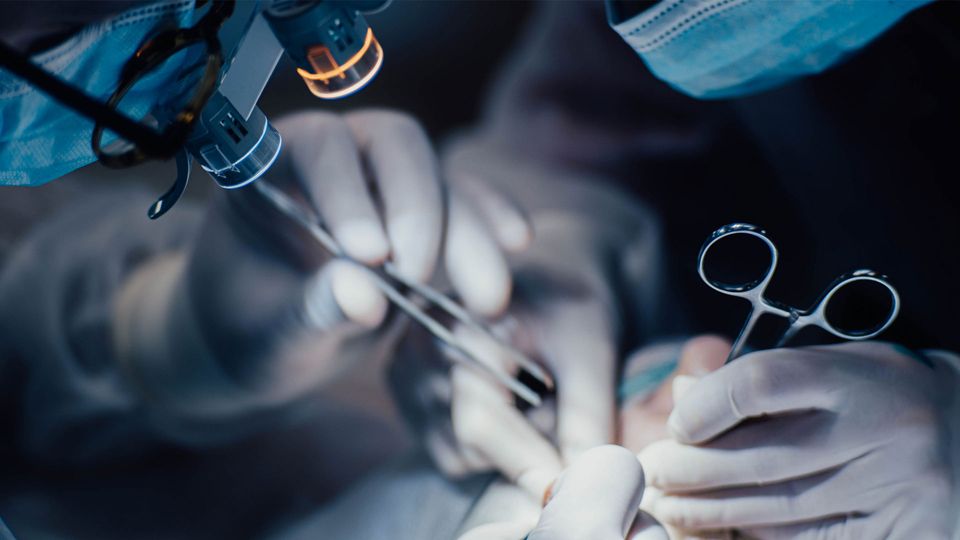
For many cancers, surgical resection is the primary treatment option. Here, the surgeon’s ability to distinguish malignant from healthy tissue is critical. Yet intraoperative decision-making often relies on frozen section histopathology – a method developed over a century ago.
While effective, it is time-consuming (taking up to 45 minutes), prone to artifacts and limited to only a handful of tissue samples per procedure. In 15–30% of cases, tissue margins that appear clear to the naked eye are later found to contain residual cancer upon post-surgical review, necessitating additional interventions.
This is where innovations in mass spectrometry (MS) – and specifically point-of-care MS platforms such as the MasSpec Pen SystemTM – are poised to reshape surgical oncology.
The missing link in point-of-care oncology
The challenge at the surgical frontier is not a lack of knowledge about cancer biomarkers – it’s the inability to deploy this knowledge in real-time. Current approaches lack the portability and speed needed to guide surgeons while a patient is still on the table.
Ambient ionization MS techniques are changing this. These methods enable near-instantaneous analysis of biomolecules directly from tissues with minimal sample preparation. The MasSpec Pen, developed by Dr. Livia Eberlin and colleagues, bridges the gap between bench and bedside by providing surgeons with a handheld, biocompatible device that interfaces directly with a mass spectrometer.
“When we developed the MasSpec Pen, the idea was to make it into a very user-friendly device that would allow surgeons, pathologists and clinicians to take advantage of the power of MS technology without requiring a PhD in analytical chemistry,” said Eberlin, a professor of surgery at Baylor College of Medicine.
In under 20 seconds, the MasSpec Pen can deliver molecular profiles that distinguish cancerous from healthy tissue, offering unprecedented support for intraoperative decisions.
“What was really lacking and that we aimed to address with the MasSpec Pen technology was a simple device between the mass spectrometer and the surgeon that was easy to use, biocompatible and was something that the surgeons could routinely employ in surgery without having training in MS. In under one minute, you can get an answer to a clinical problem that would be really challenging with other technologies,” she added.
How the MasSpec Pen works
“The MasSpec Pen allows surgeons to perform non-destructive molecular analysis and identification of tissues in vivo to identify tissues even before resection, a capability currently unavailable in surgical practice,” explained Eberlin.
The technology employs a liquid-solid extraction approach: a small droplet of water (20–50 µL) is dispensed onto tissue via the device tip, extracting metabolites and lipids that are then transferred directly to a mass spectrometer. High-resolution spectra are acquired and interpreted using machine learning algorithms such as Lasso logistic regression, which classify tissue as malignant or normal.
Key features of the MasSpec Pen include:
- Speed: Results are available in 10–20 seconds
- Non-destructive analysis: Tissue remains intact, enabling assessment even before resection.
- Ease of use: Operated by a simple foot pedal, the pen integrates seamlessly into surgical workflows.
- Accuracy: Studies report sensitivity and specificity upwards of 90% in cancers.
Importantly, the device is disposable, autoclavable and biocompatible, addressing safety and sterility concerns in the operating room.
Applications in precision oncology
The MasSpec Pen has demonstrated utility across multiple cancer types, with growing evidence supporting its clinical relevance.

The Far-Reaching Impacts of Next-Generation Sequencing
This infographic will explore how NGS has contributed to advances in personalized cancer treatment, microbiome research and environmental sciences.View Infographic
In ovarian cancer, it achieved 94.7% accuracy in distinguishing high-grade serous carcinoma from normal ovarian tissue, potentially reducing unnecessary removal of healthy tissue following chemotherapy.
“The MasSpec Pen leverages its nondestructive nature for direct and gentle analysis of tissues and the sensitivity, specificity and speed provided by MS for untargeted and accurate molecular diagnosis,” the authors of the study said.
In pancreatic cancer, the device enabled rapid intraoperative margin assessment, addressing the well-known limitations of frozen section pathology in this challenging tumor type.
“Our results provide compelling evidence that the MasSpec Pen and statistical classifiers may be valuable for assisting surgeons in making efficient, informed clinical decisions on margin status during pancreatic cancer surgery in vivo and in freshly excised tissues,” concluded the authors.
By integrating molecular data directly into surgical workflows, the MasSpec Pen exemplifies precision oncology in action – matching the right intervention to the right tissue, at the right moment.
MS beyond the operating room
The potential of MS in cancer research extends far beyond intraoperative guidance. Researchers are exploring portable MS platforms to support bedside diagnostics, real-time biopsy evaluation and monitoring treatment response in outpatient settings.
Recent developments in miniaturized MS systems – such as compact Orbitrap and time-of-flight instruments – are making it increasingly feasible to deploy molecular diagnostics in decentralized healthcare environments.

Changing the Landscape of Screening: AI-Powered ASMS Ligand Identification
This webinar explores how advanced screening technologies strengthen affinity selection mass spectrometry (ASMS) workflows to speed up timelines, improve precision and uncover novel binders.View Webinar
In oncology, this progress could enable rapid tumor profiling at the bedside or in outpatient clinics, reducing delays in treatment planning. Further, it could allow for real-time monitoring of circulating tumor metabolites to assess therapy effectiveness or detect early signs of relapse and point-of-care molecular screening in low-resource settings, helping to expand global access to precision oncology.
Integrating MS with artificial intelligence and cloud-based data sharing is also emerging as a powerful strategy. Real-time spectral analysis combined with machine learning could provide automated clinical decision support, while networked platforms would allow remote expert consultation for complex cases.
Challenges and path to clinical translation
Despite these advances, barriers remain. High acquisition costs, large instrument size and the need for specialized operators continue to limit the widespread deployment of MS in hospitals.
However, efforts are underway to overcome these challenges through the development of fully integrated point-of-care MS devices that combine handheld sampling tools with portable MS units. Additional strategies include the creation of simplified user interfaces that enable non-specialists, such as surgeons or nurses, to operate the tools with minimal training, as well as the regulatory validation and standardization of MS-based diagnostic workflows to meet clinical certification requirements.
Miniaturization and portability are at the heart of MS’s clinical future. Compact platforms, coupled with user-friendly interfaces like the MasSpec Pen, could democratize molecular diagnostics, moving them from specialized labs to point-of-care settings.
For oncology, this means faster diagnoses, fewer repeat surgeries and improved patient outcomes. Beyond surgery, handheld MS devices could support bedside cancer diagnostics, guide biopsies or even monitor treatment response in real-time.
The MasSpec Pen is not just an incremental advance – it represents a paradigm shift in intraoperative cancer care. By delivering MS’s molecular power into the hands of surgeons, it enables precise, rapid and minimally invasive tissue characterization at the point-of-care.


